Ayurveda has a unique approach to diagnosis, which is holistic and based on observing the patient’s physical, mental, and emotional state. The main diagnostic methods in Ayurveda are collectively known as "Ashta Vidha Pariksha" (Eightfold Examination) and "Trividha Pariksha" (Threefold Examination).
Tonometry and Optical Coherence Tomography (OCT) – Advanced Clinical Discussion
Both Tonometry and Optical Coherence Tomography (OCT) are crucial diagnostic tools in ophthalmology, primarily used for glaucoma assessment, retinal diseases, and corneal evaluations.
I. Tonometry – Measuring Intraocular Pressure (IOP)
1. What is Tonometry?
Tonometry is a diagnostic test used to measure intraocular pressure (IOP), which is the pressure exerted by the aqueous humor inside the eye. Abnormal IOP can lead to conditions like glaucoma, which may cause irreversible blindness.
2. Normal and Abnormal IOP Values
Condition
IOP (mmHg)
Clinical Relevance
Normal Eye Pressure
10-21 mmHg
No risk of glaucoma
Ocular Hypertension
> 21 mmHg
Risk of glaucoma
Glaucoma
> 24 mmHg
Optic nerve damage possible
Hypotony (Low IOP)
< 6 mmHg
Risk of retinal detachment
3. Types of Tonometry Methods
Type
Principle
Procedure
Clinical Use
Goldmann Applanation Tonometry (GAT)
Measures force needed to flatten the cornea
Fluorescein dye + slit lamp
Gold standard for IOP measurement
Non-Contact (Air-Puff) Tonometry
Uses a puff of air to flatten the cornea
No contact with eye
Quick, but less accurate
Rebound Tonometry (iCare)
Measures the rebound of a probe from cornea
No anesthesia required
Used in children and home monitoring
Dynamic Contour Tonometry (DCT)
Measures IOP independent of corneal thickness
Uses a special probe
More accurate than GAT
Schiøtz Indentation Tonometry
Indents the cornea to measure IOP
Less commonly used
Useful in post-surgical cases
4. Clinical Applications of Tonometry
Glaucoma Diagnosis & Monitoring
Post-Surgical IOP Monitoring
Screening for Ocular Hypertension
Assessing IOP Fluctuations in Trauma Cases
II. Optical Coherence Tomography (OCT) – Retinal & Corneal Imaging
1. What is OCT?
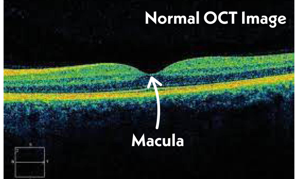
OCT is a non-invasive imaging technique that provides high-resolution cross-sectional images of the retina, optic nerve, and cornea. It works on the principle of interferometry using infrared light to create detailed 3D images.
2. Clinical Applications of OCT
Disease
OCT Findings
Clinical Importance
Glaucoma
Retinal nerve fiber layer (RNFL) thinning
Early detection
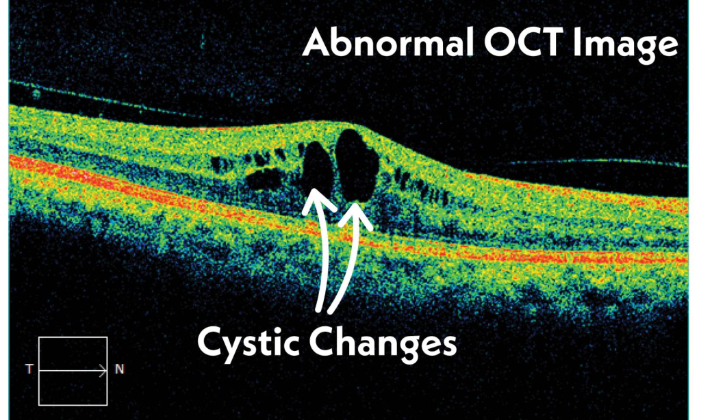
Diabetic Retinopathy
Macular edema, hemorrhages
Detects fluid leakage
Age-Related Macular Degeneration (AMD)
Drusen, subretinal fluid
Helps guide treatment
Retinal Detachment
Retinal separation from RPE
Urgent surgical referral
Keratoconus
Corneal thinning, irregularity
Pre-LASIK screening
Optic Neuritis (MS-related)
RNFL thinning
Neuro-ophthalmic evaluation
3. Types of OCT and Their Uses
OCT Type
Primary Use
Details
Spectral-Domain OCT (SD-OCT)
Retina & macula imaging
Higher speed, high resolution
Swept-Source OCT (SS-OCT)
Deeper tissue imaging
Ideal for choroidal & optic nerve analysis
Anterior Segment OCT (AS-OCT)
Cornea & anterior chamber
Used for keratoconus & glaucoma
OCT Angiography (OCTA)
Vascular imaging
Detects diabetic & macular vascular diseases
4. How to Perform OCT?
✅ Step 1: The patient places their chin on a rest and focuses on a fixation light. ✅ Step 2: The machine scans the retina using infrared light, capturing images within seconds. ✅ Step 3: The software processes the images and generates 3D cross-sectional views. ✅ Step 4: The ophthalmologist analyzes the thickness of retinal layers, optic nerve health, and presence of fluid leakage.
III. Tonometry vs. OCT – Comparison Table
Parameter
Tonometry
OCT
Purpose
Measures intraocular pressure (IOP)
High-resolution retinal imaging
Clinical Use
Glaucoma detection & monitoring
Retinal, optic nerve, and corneal disease
Procedure
Applanation (contact) or air-puff (non-contact)
Infrared scanning (non-invasive)
Key Indicator
Elevated IOP suggests glaucoma
Retinal nerve fiber layer thinning in glaucoma
Limitations
Affected by corneal thickness
Cannot visualize deep optic nerve damage
Advanced Clinical Discussion on Optical Coherence Tomography (OCT)
Introduction
Optical Coherence Tomography (OCT) is a non-invasive imaging technique that provides high-resolution cross-sectional images of the retina, macula, optic nerve, and cornea. It is widely used in ophthalmology, neurology, and vascular medicine for early diagnosis and monitoring of various conditions.
I. Principles of OCT
Works on the principle of low-coherence interferometry using near-infrared light (800-1300 nm).
Measures light reflections from different tissue layers to create a high-resolution 3D image.
Provides micron-level (1-15 μm) resolution, superior to ultrasound.
Comparison of Imaging Modalities:
Modality
Resolution
Penetration
Used for
Ultrasound (USG)
150 μm
Deep structures
Retinal detachment, tumors
Fundus Photography
20-50 μm
Surface imaging
Diabetic retinopathy screening
Fluorescein Angiography (FA)
20-30 μm
Blood vessel imaging
Vascular diseases
OCT
1-15 μm
Limited to retina & cornea
Glaucoma, AMD, diabetic macular edema
II. Types of OCT and Their Clinical Applications
OCT Type
Clinical Use
Key Features
Spectral-Domain OCT (SD-OCT)
Retinal diseases
High speed, high resolution
Swept-Source OCT (SS-OCT)
Choroidal imaging
Better depth penetration
OCT Angiography (OCTA)
Vascular diseases
Non-invasive, detects ischemia
Anterior Segment OCT (AS-OCT)
Cornea, angle evaluation
Useful for keratoconus, glaucoma
III. Clinical Interpretation of OCT in Different Diseases
Ganglion Cell Layer Analysis – Early sign of neurodegeneration in MS
VII. Limitations of OCT
🔹 Cannot penetrate highly pigmented tissues (e.g., retinal hemorrhage) 🔹 Limited field of view compared to wide-field fundus photography 🔹 Artifacts due to media opacity (cataracts, corneal edema)